- Home
- About us
- Laboratories
- Central Nervous System Physiology
- Central Nervous System functions compensation Physiology
- Immunology and Tissue Engineering
- Smooth Muscle Physiology
- Toxinology and Molecular Systematics
- Sensorimotor Integration
- Laboratory of Histochemistry and Morphology
- Human Psychophysiology
- Integrative Biology
- Purification, Certification and Standardization of Physiologically active substances
- Neuroendocrine Relationships
- Distance Լearning Center
- Laboratory of Hyperspectral Imaging of Surgical Targets
- Biophysics Lab
- News & Events
- Master's Program
- Research
- Councils
- Contact Us
- OIPH Docs
L. A. Orbeli Institute of
Physiology NAS RA
Laboratory of Histochemistry and Morphology

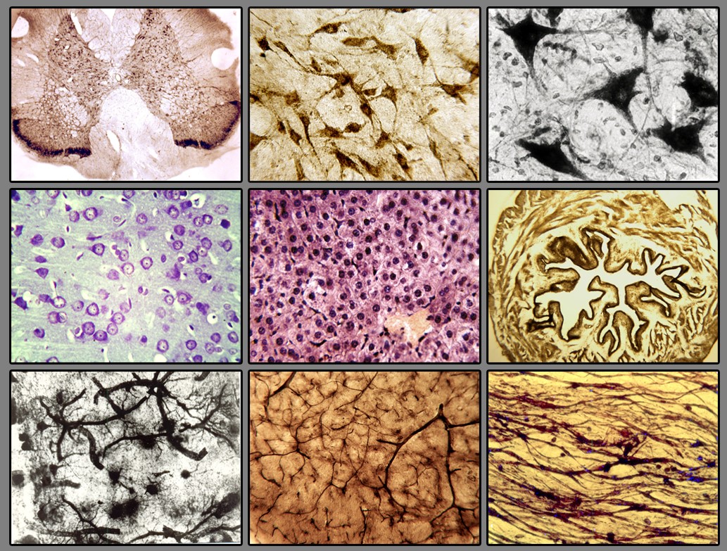
The objective of the laboratory is to investigate the morphological aspects of pathological, restorative, and protective processes in various tissues to provide a basis for the implementation of different therapeutic agents in clinical practice.
Scientific research tasks include conducting preclinical animal experiments under normal conditions and with various pathologies. The laboratory conducts studies on the effects of different biologically active substances, toxins from various snake species, and animal-derived biogenic neuromodulators. These substances can protect or regenerate the damaged cellular components. The aim of our lab is to investigate the mechanisms of their action and develop next-generation medicinal preparations for the prevention and treatment of non-specific and specific neurodegenerative diseases.

The scientific interests of the laboratory encompass the following areas: 1. Neurodegenerative diseases in humans and animals (such as Alzheimer's disease and Parkinson's disease) 2. Metabolic diseases in humans and animals (diabetes etc.), alcoholism; 3. Study of bioactive compounds with vasodilator and anticonvulsant activities.
The morphological studies conducted in the laboratory are comprehensive in nature and are performed at a modern level using techniques such as light microscopy, histochemistry, histomorphometry, and various histological methods. The obtained data open new possibilities for further investigation of the morphofunctional state of cellular structures in the brain, spinal cord, and various organs under both normal and pathological conditions. These findings are of particular interest in practical medicine, especially for elucidating the role and influence of bioactive substances.
The Histochemistry and Morphology Laboratory offers services to schools, colleges, institutes, medical universities, and research organizations for educational and research purposes Laboratory's Material and Technical Equipment: • Dissection and preparation of biological tissue samples, production of paraffin blocks, and preparation of histological slides from various tissues for examination by light microscopy. • Light microscopy of histological slides using an OPTON (Germany) light microscope equipped with Amscope MU800 8 MP (USA) digital camera and ToupTek ToupView software. • Digital input, analysis, and archiving of full-color images. • Morphometry using ImageJ software. The laboratory is equipped with a wide range of high-quality equipment and instruments to perform the aforementioned tasks. Tissue samples can be processed, embedded in paraffin, and sectioned for histological examination. The OPTON light microscope, equipped with an Amscope MU800 digital camera, allows for the visualization and capture of high-resolution images. The ToupTek ToupView software enables image analysis and facilitates the digital input, processing, and storage of color images. Additionally, morphometric studies can be conducted using ImageJ for quantitative measurements and data analysis.
| Publications |
|---|
12. Sarkisyan SH, Danielyan MH, Darbinyan LV, Simonyan KV, Chavushyan VA. The effects of vibration on the neuronal activity of lateral vestibular nuclei in unilaterally labyrinthectomized rats. Brain Struct Funct. 228:463–473(2023). https://doi.org/10.1007/s00429-022-02588-6
13. Chavushyan VA, Simonyan KV, Danielyan MH, Avetisyan LG, Darbinyan LV, Isoyan AS, Lorikyan AG, Hovhannisyan LE, Babakhanyan MA, Sukiasyan LM. Pathology and prevention of brain microvascular and neuronal dysfunction induced by a high-fructose diet in rats. Metab Brain Dis. 2023 Jan; 38(1):269-286. https://doi.org/10.1007/s11011-022- 01098-y
14. Yenkoyan K., Margaryan T., Matinyan S., Chavushyan V., Danielyan M., Davtyan T., Aghajanov M. Effects of β-amyloid (1-42) Administration on the Main Neurogenic Niches of the Adult Brain: Amyloid-Induced Neurodegeneration Influences Neurogenesis. И https://doi.org/10.3390/ijms232315444
15. Simonyan R.M., Simonyan K.V., Simonyan G.M., Khachatryan H.S., Babayan M.A., Danielyan M.H., Darbinyan L.V., Simonyan M.A. Superoxide-producing thermostable associate from the small intestines of control and alloxan-induced diabetic rats: quantitative and qualitative changes. BMC Endocr Disord., 22(1), Article number: 250 (2022), p. 1-6. https://doi.org/10.1186/s12902-022-01160-x
16. Danielyan MH, Karapetyan KV, Nebogova KA, Nazaryan O., Khachatryan VP. Effects of Galarmin and Cobra Venom on the Morphofunctional State of the Substantia Nigra in a Rat Model of Parkinson's Disease. Neurophysiology, 53(1): 22-29 (2021). https://doi.org/10.1007/s11062-021-09909-1
17. Aghajanov M, Matinyan S, Chavushyan V, Danielyan M, Karapetyan G, Mirumyan M, Fereshetyan K, Harutyunyan H, Yenkoyan K. The Involvement of Insulin-Like Growth Factor 1 and Nerve Growth Factor in Alzheimer’s Disease-Like Pathology and Survival Role of the Mix of Embryonic Proteoglycans: Electrophysiological Fingerprint, Structural Changes and Regulatory Effects on Neurotrophins. International Journal of Molecular Sciences, 22(13):7084 (2021). https://doi.org/10.3390/ijms22137084 .
18. Fereshetyan K., Chavushyan V., Danielyan M., Yenkoyan K. Assessment of behavioral, morphological and electrophysiological changes in prenatal and postnatal valproate induced rat models of autism spectrum disorder. Scientific Reports, 11, 23471:1-17(2021). https://doi.org/10.1038/s41598-021-02994-6
19. Chavushyan V, Matinyan S, Danielyan M, Aghajanov M, Yenkoyan K. Embryonic proteoglycans regulate monoamines in the rat frontal cortex and hippocampus in Alzheimer’s disease-like pathology. Neurochemistry International, 140, 104838 (2020) https://doi.org/10.1016/j.neuint.2020.104838
20. Aghajanov M, Chavushyan V, Matinyan S., Danielyan M, Yenkoyan K. Alzheimer’s disease-like pathology-triggered oxidative stress, alterations in monoamines levels, and structural damage of locus coeruleus neurons are partially recovered by a mix of proteoglycans of embryonic genesis. Neurochemistry International, 131, 104531 (2019). https://doi.org/10.1016/j.neuint.2019.104531
21. Kazaryan K.V., Danielyan M.A., Chibukhchyan R.G., M argaryan Sh.G. Histamine-Mediated Regulation of Electrical Activity during the Bladder–Urethra Interaction in Rats. Journal of Evolutionary Biochemistry and Physiology, 54(1):50-58(2018). https://doi.org/10.1134/S0022093018010064
22. Ghazaryan N.A., Simonyan K.V., Danielyan M.H., Zakaryan N.A., Ghulikyan L.A., Kirakosyan G.R., Chavushyan V.A., Ayvazyan N.M. Neuroprotective Effects of Macrovipera lebetina Snake Venom in the Model of Alzheimer’s Disease. Neurophysiology, 49(6): 412-423 (2017). https://doi.org/10.1007/s11062-018-9704-8
Group members


